Walnut CT
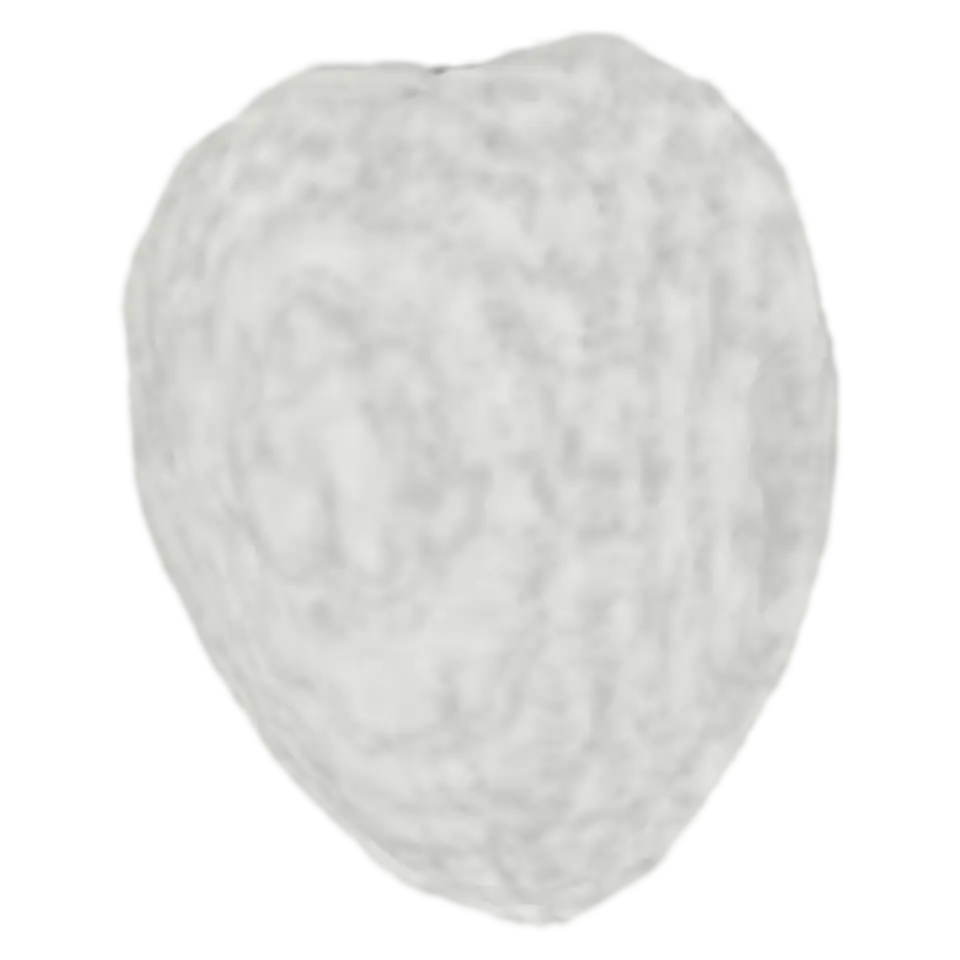
This is a real walnut, scanned with X-ray CT at CWI in Amsterdam: more than a thousand X-ray pictures taken from all around it, worked back into a volume of how dense the walnut is at every point.
This picture is a cloud of paint. You can , , , , , , and .
Paint with your finger. It stays on these splats, wherever they go next.
Your paint is on this cloud. to see the sharp picture.
- Shelf
- Imaging
- Kind
- Built from a recipe
- Made from
- Drag up or down on the walnut to cut into it.
- Sound
- Yes
- Licenses
- CC BY 4.0
How to play
Drag up or down on the walnut to cut into it. Tap to see only its shell.
Its controls
| Control | Kind | Range | Start |
|---|---|---|---|
| Shell only, or the whole walnut | Tap | ||
| Shell only | Switch | Off | |
| Cut | List | 3 choices | Front to back |
| Colors | List | 4 choices | CT gray |
About it
This is a real walnut, scanned with X-ray CT at CWI in Amsterdam: more than a thousand X-ray pictures taken from all around it, worked back into a volume of how dense the walnut is at every point. Each splat here is one small block of that volume, 0.3 mm across, bright where the walnut is dense.
Drag up or down to cut into it and see the woody shell, the kernel's folded lobes and the air between them. Choose the cut's direction and the colors (CT gray, warm, night vision or infrared) in the Splash tab. Tap to show only the densest parts: the kernel melts away and the shell stays. The scan is Walnut 1 of a dataset by Der Sarkissian, Lucka, van Eijnatten, Colacicco, Coban and Batenburg (CC BY 4.0).
Sources and credits
| Work | By | License |
|---|---|---|
| Cone-Beam X-Ray CT Data Collection Designed for Machine Learning: Samples 1-8 (Walnut 1) | Henri Der Sarkissian, Felix Lucka, Maureen van Eijnatten, Giulia Colacicco, Sophia Bethany Coban, K. Joost Batenburg (CWI) | CC BY 4.0 |
In The Splash Manual
Share it
This link opens it in the Playground. To put it on your own page, add the element and one script tag. Every option.
https://splashery.app/play/?toy=walnut-ct
<script type="module" src="https://splashery.app/play/src/element.js"></script>
<splashery-toy toy="walnut-ct"></splashery-toy>
Nearby
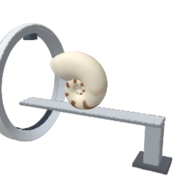 CT ExplainedA CT scanner (computed tomography) turns an X-ray tube and a curved row of detectors around the object, taking hundreds of views from every side.
 Fruit MRIAn MRI scanner (magnetic resonance imaging) uses no X-rays.
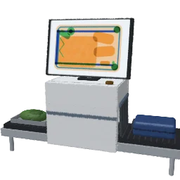 Airport X-rayAn airport scanner sends a fan of X-rays through each bag as the belt carries it past, and a row of detectors measures what gets through, one line at a time.
 Electron MicroscopeA scanning electron microscope sweeps a fine beam of electrons across a sample in a vacuum.